Livedoid Vasculopathy: What It Looks Like and How to Manage It
Here’s a blunt fact: livedoid vasculopathy often gets mistaken for ordinary leg ulcers, but it’s driven by tiny blood clots in the skin’s small vessels. That matters because treating it like a simple skin wound usually fails. This quick guide tells you how to spot it, what doctors test for, and concrete steps that actually help.
Signs you shouldn’t ignore: recurring painful red or purple net-like patches (livedo), small atrophie blanche scars, and slow-healing ulcers on the lower legs and ankles. Pain is often out of proportion to what the wound looks like. Lesions can come and go, and scarring is common after they heal.
What causes it? Livedoid vasculopathy is mainly a thrombotic (clotting) problem in tiny skin vessels. Doctors often look for blood-clotting abnormalities—factor V Leiden, antiphospholipid antibodies, elevated homocysteine, or other hypercoagulable states. It can appear on its own or alongside autoimmune issues, but inflammation in the usual sense is not the main driver.
Tangible steps for diagnosis
Ask for a skin biopsy when ulcers are unexplained—histology shows fibrin plugs and vessel wall thrombosis. Blood tests should check clotting factors, antiphospholipid panel, basic autoimmune markers (ANA), and glucose/lipid levels. A vascular exam rules out arterial disease; ankle-brachial index or Doppler studies help. Getting a dermatologist and hematologist involved early speeds correct diagnosis.
Treatment and wound care that help
Treatment focuses on preventing further microclots and supporting healing. Anticoagulation is often central—doctors may use warfarin or direct oral anticoagulants under specialist guidance. Low-dose aspirin or other antiplatelet drugs are sometimes added. When an autoimmune cause is found, targeted immunotherapy (for example, IVIG or other agents) might be used, but that’s decided case-by-case.
Local wound care matters: keep ulcers clean, use moist dressings, and change them per clinic advice. Avoid aggressive debridement unless guided by a specialist. Compression stockings can help if venous disease is also present, but only after vascular assessment. Control smoking, high blood sugar, and high cholesterol—those raise clot risk and slow healing.
Pain control and infection prevention are practical priorities. Topical or short-term oral antibiotics only if infection is present. For persistent cases, some clinics offer pentoxifylline to improve microcirculation or prostacyclin-like therapies; these are added by specialists when needed.
When to see a doctor: get urgent care if ulcers suddenly swell, produce heavy discharge, develop fever, or pain spikes. If wounds linger beyond a few weeks despite basic care, push for biopsy and hematology input. With proper treatment most people improve, though relapses and scarring can occur.
Bottom line: livedoid vasculopathy is a small-vessel clotting disorder that looks like ordinary leg ulcers but needs different care. Early biopsy, clotting tests, specialist input, and targeted anticoagulation plus careful wound management give the best chance to heal and prevent new ulcers.
The potential benefits of betamethasone for treating livedoid vasculopathy
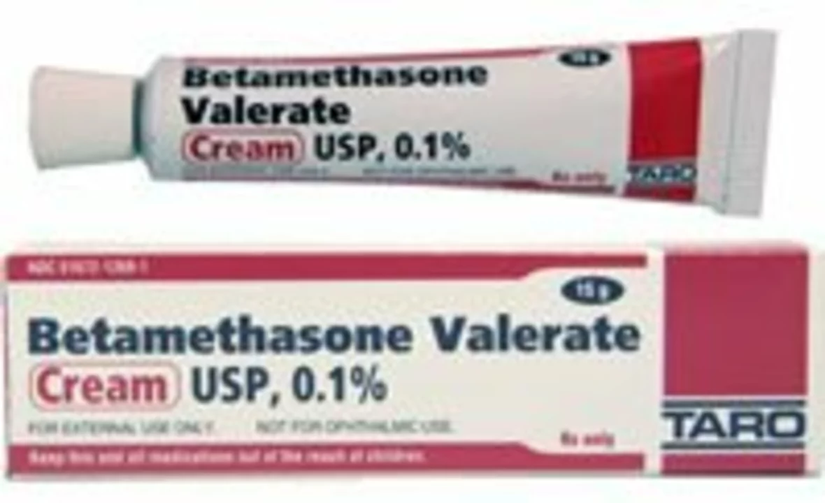
In my recent research, I've discovered the potential benefits of betamethasone for treating livedoid vasculopathy. Livedoid vasculopathy is a rare and painful skin condition where blood vessels become inflamed, causing ulcers and discoloration. Betamethasone, a powerful corticosteroid, has been found to reduce inflammation and promote healing in affected areas. With its anti-inflammatory and immunosuppressive properties, betamethasone offers a promising treatment option for those suffering from livedoid vasculopathy. I'm eager to follow any upcoming research and clinical trials to better understand the full potential of this treatment.
© 2026. All rights reserved.

